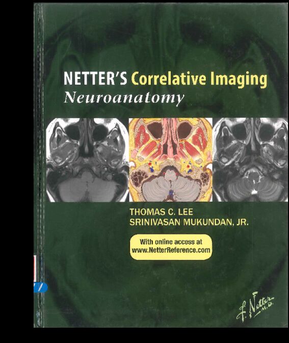
书名:Netter’s correlative imaging. Neuroanatomy
责任者:Thomas C. Lee | Srinivasan Mukundan | Jr. ; Frank H. Netter | contributing illustrators | Tiffany Slaybaugh DaVanzo | Carlos Machado. | Netter, Frank H.
出版时间:2015
出版社:Elsevier/Saunders
前言
The study of anatomy, by its very nature, exposes the student to both the forms and the functions responsible for the inner workings of the human body—one of the most elegant demonstrations of the synthesis of art and science. This statement is never truer than when reviewing the works of Frank H. Netter, MD (1906-1991). His monumental works of art have educated countless generations of physicians and will continue to do so for many years to come
The evolution of cross-sectional imaging technologies, including computed tomography (CT) and magnetic resonance imaging (MRI), has allowed physicians and aspiring physicians the ability to peer into the human body in many ways previously impossible. This, in turn, has forced the review of anatomy in a manner that was not contemplated when Dr. Netter began rendering his artwork more than three-quarters of a century ago. Limited spatial and temporal resolution, tissue parameters, image contrast, and partial volume averaging are all issues that confound the interpretation of MRI and CT images and are not part of conventional anatomic teaching in the dissection laboratory.
Despite the changing nature of the anatomy education problem, Dr. Netter's approach still provides the template for how to teach modern medical imaging. By directly correlating cross-sectional medical images with "Netter art" that is voxel matched, the student is provided with the key to unlock the anatomy hidden within.
Many existing textbooks with radiology images and clarifying illustrations focus on showing 3D renderings for better understanding of the entire course of a particular structure, such as a cranial nerve. Our goal, however, is to provide a more rigorous "slicc-by-slice" reference guide for both the cross-sectional medical image and the corresponding illustration. For instance, someone may understand the general course of the facia! nerve, yet still have difficulty identifying the fractional component of this structure on a single axial image. Conversely, individuals may need help identifying an unknown structure on a given image, particularly in the coronal or sagittal planes. Finally, the imaging characteristics on CT versus different sequences of MRI, such as T1- or T2-weighted imaging, can often be confusing and will hopefully be partially clarified through this book.
This volume remains true to the goals of the Netter's Correlative Imaging series. There is high-quality imaging, allowing the demonstration of important anatomic structures, including structures not always included in other sources. The book also serves as a user-friendly anatomy reference for commonly employed imaging techniques of the brain, head, neck, and spine. As with the other volumes in the series, the text is not inclusive of pathology, In addition, normal variant anatomy and clinical pearls are presented.
Structures are labeled using the most common terms and should be acceptable to radiologists, neurosurgeons, and neurologists.
It is our hope that this volume will serve as a primary source of knowledge to the novice and as a reference text for the seasoned veteran In either case, we hope that it will be useful on a daily basis.
查看更多
目录
PART 1 BRAIN
1 OVERVIEW OF BRAIN 2
2 BRAIN 15
Axial, 16-61
Coronal, 62-97
Sagittal, 98-123
3 THALAMUS AND BASAL GANGLIA 125
Axial, 126-135
Coronal, 136-145
4 LIMBIC SYSTEM 147
Axial, 148-155
Coronal, 156-167
Sagittal, 168-173
5 BRAINSTEM AND CRANIAL NERVES 175
Olfactory Nerve (CN I)
Axial, 176-177
Coronal, 178-179
Optic Nerve (CN II)
Axial, 180-185
Coronal, 186-197
Sagittal, 198-199
Oculomotor Nerve (CN III)
Axial, 200-207
Coronal, 208-217
Trochlear Nerve (CN IV)
Axial, 218-221
Coronal, 222-223
Trigeminal Nerve (CN V)
Axial, 224-241
Sagittal, 242-243
Coronal, 244-245
Abducens (CN VI), Facial (CN VII), and Vestibulocochlear (VIII) Nerves
Axial, 246-259
Sagittal, 260-263
Glossopharyngeal (CN IX), Vagus (CN X), Accessory (CN XI), and Hypoglossal (CN XII) Nerves
Axial, 264-271
6 VENTRICLES AND CEREBROSPINAL FLUID CISTERNS 273
Axial, 274-285
Coronal, 286-291
Sagittal, 292-293
7 SELLATURCICA 295
Coronal, 296-303
Sagittal, 304-305
PART 2
8 OVERVIEW OF HEAD AND NECK 308
9 PARANASAL SINUSES 321
Axial, 322-337
Coronal, 338-353
10 ORBITS 355
Axial, 356-367
Coronal, 368-379
11 MANDIBLE AND MUSCLES OF MASTICATION 381
Axial, 382-389
Coronal, 390-397
12 TEMPORAL BONE (MIDDLE EAR, COCHLEA, VESTIBULAR SYSTEM) 399
Axial, 400-411
Coronal, 412-419
13 ORAL CAVITY, PHARYNX, AND SUPRAHYOID NECK 421
Axial, 422-437
Coronal, 438-453
Sagittal, 454-469
14 HYPOPHARYNX, LARYNX, AND INFRAHYOID NECK 471
Axial, 472-487
Coronal, 488-503
Sagittal, 504-519
PART 3
15 OVERVIEW OF SPINE 523
16 SPINE 535
Cervical Spine
Axial, 536-551
Coronal, 552-561
Sagittal, 562-571
Thoracic Spine
Axial, 572-577
Sagittal, 578-583
Lumbosacral Spine
Axial, 584-595
Sagittal, 596-605
查看PDF
查看更多
馆藏单位
中国医科院医学信息研究所